The science
Why ILUMI pairs blue light with red light
Whitening isn't bleach painted onto a tooth. It's a controlled oxidation reaction inside enamel. Light is a separate question, and we try to be straight about what the research does and does not show.
Peroxide breaks the stain, not the tooth
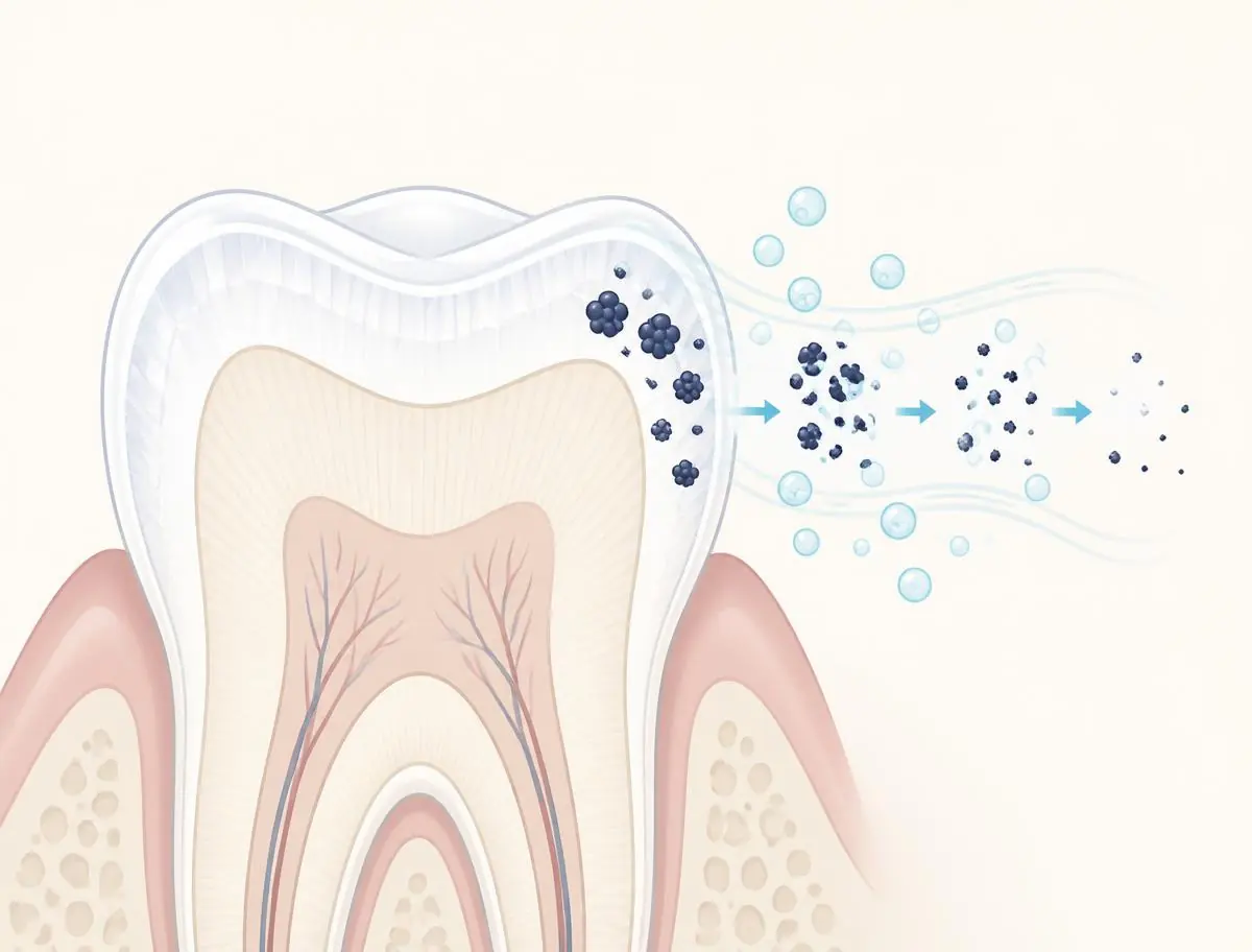
The color of a stained tooth comes from chromogens — large, tightly conjugated organic molecules from coffee, tea, wine, tobacco and food pigments that lodge in the porous structure of enamel and dentin.
Carbamide peroxide breaks down into hydrogen peroxide, which in turn releases reactive oxygen species. Those radicals cleave the double bonds that make chromogens absorb visible light. The molecules aren't scrubbed away — they're broken into smaller, lighter-colored fragments that reflect rather than absorb. The mineral structure of the enamel itself is not the target of the reaction.
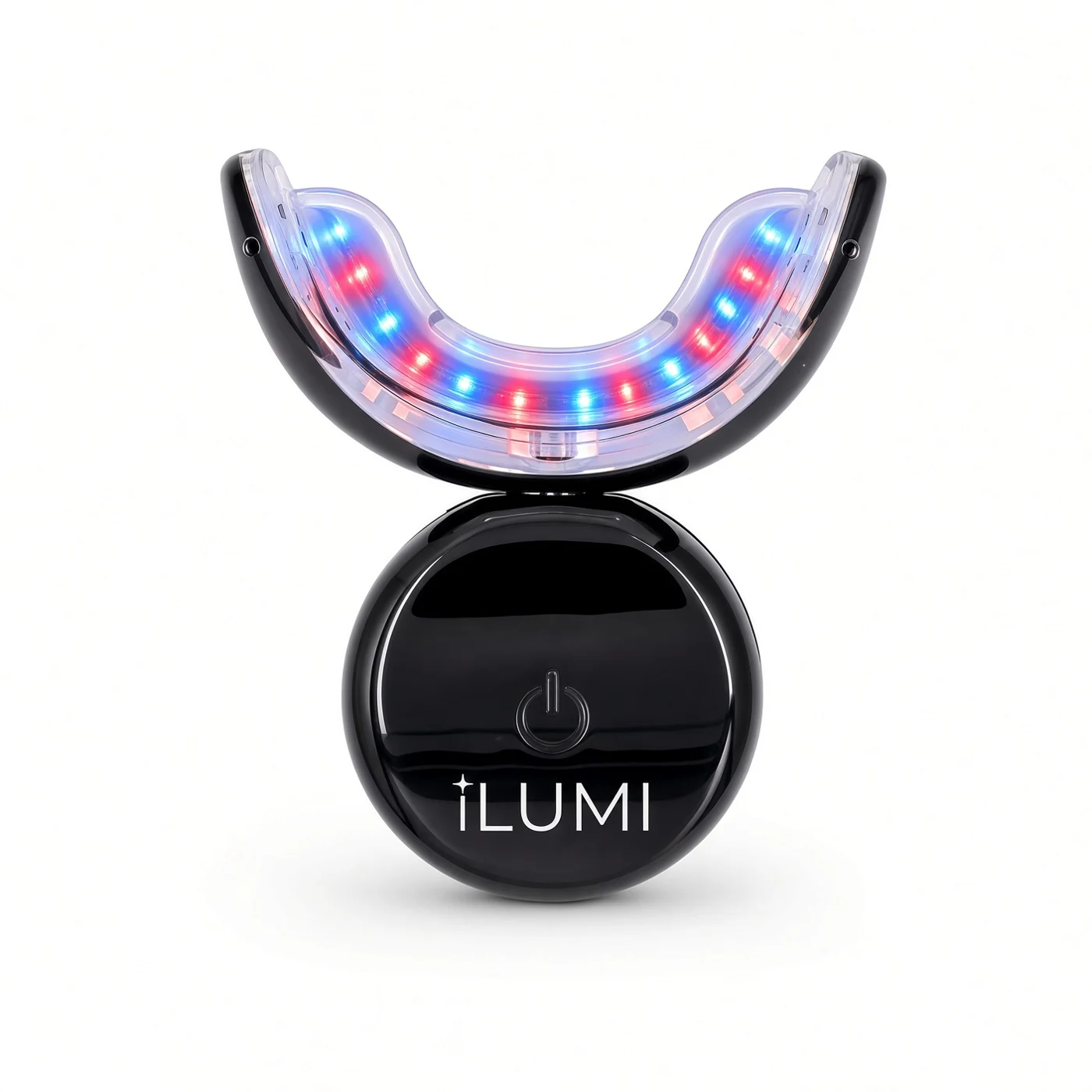
Blue light goes with the gel. Red light is there for comfort.
Most whitening lights use one wavelength. ILUMI uses two, because whitening is only useful if you actually finish the round, and sensitivity is a common reason people stop. The blue band is the one used with the gel; the red band is the one that has been studied for easing sensitivity after whitening.
Blue — roughly 450–480 nm
Blue light in this band is the wavelength used with the gel in the ILUMI mouthpiece. To be straightforward: systematic reviews of in-office whitening have not found that light activation improves the final result, so we do not claim that it does. What the device adds is a timed, hands-free 10-minute session.
Red — roughly 620–660 nm
Red light is the wavelength band used in photobiomodulation. A 2026 umbrella review of systematic reviews found that photobiomodulation reduced post-whitening sensitivity in placebo-controlled trials without reducing whitening. Those were in-office protocols with specific settings, so the result may not carry over to every device. We include red light because sensitivity is a common reason people stop. It runs at the same time as the blue.
Both lights run together for one 10-minute session, and the device switches itself off. The kit also includes a blue desensitizing gel pen. Results and comfort vary from person to person, and you can come back for touch-ups whenever coffee, wine, or life starts to dull your smile again.
The formula is where sensitivity is won or lost
Concentration, chosen deliberately
Sensitivity scales with peroxide concentration and contact time. The gel is formulated to sit in a band that still moves shade while aiming to go easy on sensitive teeth.
pH-balanced
An acidic gel can demineralise enamel and make teeth feel rough and reactive. The ILUMI gel is pH-balanced and made in an FDA-registered facility.
A tray that keeps gel off gums
Most 'chemical burn' complaints about whitening are gel migrating onto soft tissue. Correct tray geometry solves a problem no formula change can.
From the formulator
"I spend my working life inside teeth. I built this system because I wanted a whitening protocol I could hand a patient without a disclaimer — one that moves shade quickly, respects enamel, and doesn't punish people with sensitivity three days in. Every choice here, the concentration, the pH, the two wavelengths, exists to serve those three things at once."
Dr. Dave Steuer, D.M.D., Ph.D. — Harvard-trained endodontist, Ph.D. Natural Health and Medicine, Ph.D. Sports Nutrition, formulator of ILUMI
The limit of the chemistry
Why whitening does nothing to a crown, veneer or filling
Everything on this page depends on one property of natural teeth: enamel and dentin are porous, so peroxide can diffuse in and oxidize the pigment molecules trapped there. Porcelain, ceramic, zirconia and composite resin have no such porosity and no chromogens to break. Peroxide simply washes over the surface. A crown that was shade A2 the day it was cemented is still shade A2 after ten whitening sessions.
That has a practical consequence people rarely think about until it's too late. The natural teeth around the restoration keep lightening. A crown that used to disappear into your smile can end up reading dark or yellow next to the teeth beside it — and unlike a stain, that mismatch does not fade. The only fix is to have the restoration remade to the new shade.
The sequence that avoids the problem: whiten first, let the shade stabilize for about two weeks (freshly whitened enamel rebounds slightly and rehydrates), then have your dentist match any crown, veneer or visible bonding to where your teeth actually landed. Tooth-colored fillings on front teeth deserve the same check — they were matched to a darker shade at the time they were placed. If most of your visible smile is restorations, whitening will change very little about how it looks, and that's worth knowing before you buy.
Lightens
Natural enamel and dentin — extrinsic coffee, tea, wine, tobacco and food staining most of all; intrinsic discoloration more slowly, over repeated rounds.
Does not lighten
Porcelain and ceramic crowns, zirconia, veneers, composite bonding and fillings, bridge units and implant crowns. Also enamel hidden under fixed braces brackets.
Before the stain
Your teeth are already wearing a film
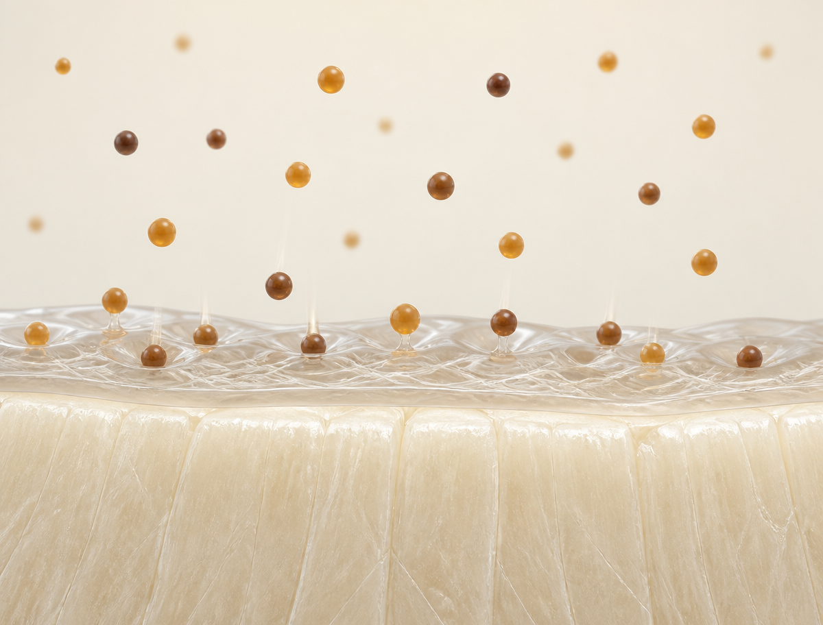
Within minutes of a professional cleaning, proteins from saliva adsorb onto enamel and form the acquired enamel pellicle — a thin protein layer that reforms continuously and cannot be brushed away for long. This film is genuinely protective: it lubricates the tooth surface, buffers acid, and limits mineral loss. It is also the surface that pigment actually binds to. Chromogens from coffee, tea, red wine and tobacco largely attach to the pellicle rather than directly to enamel.
That has a practical consequence worth understanding before you whiten: the film reforms within hours of any cleaning, so new staining begins again immediately. Whitening resets where your color sits; it does not stop the surface from re-accumulating pigment. Maintenance is not a marketing idea — it is what the biology requires.
The living stain
Some discoloration isn't a stain at all — it's an ecosystem
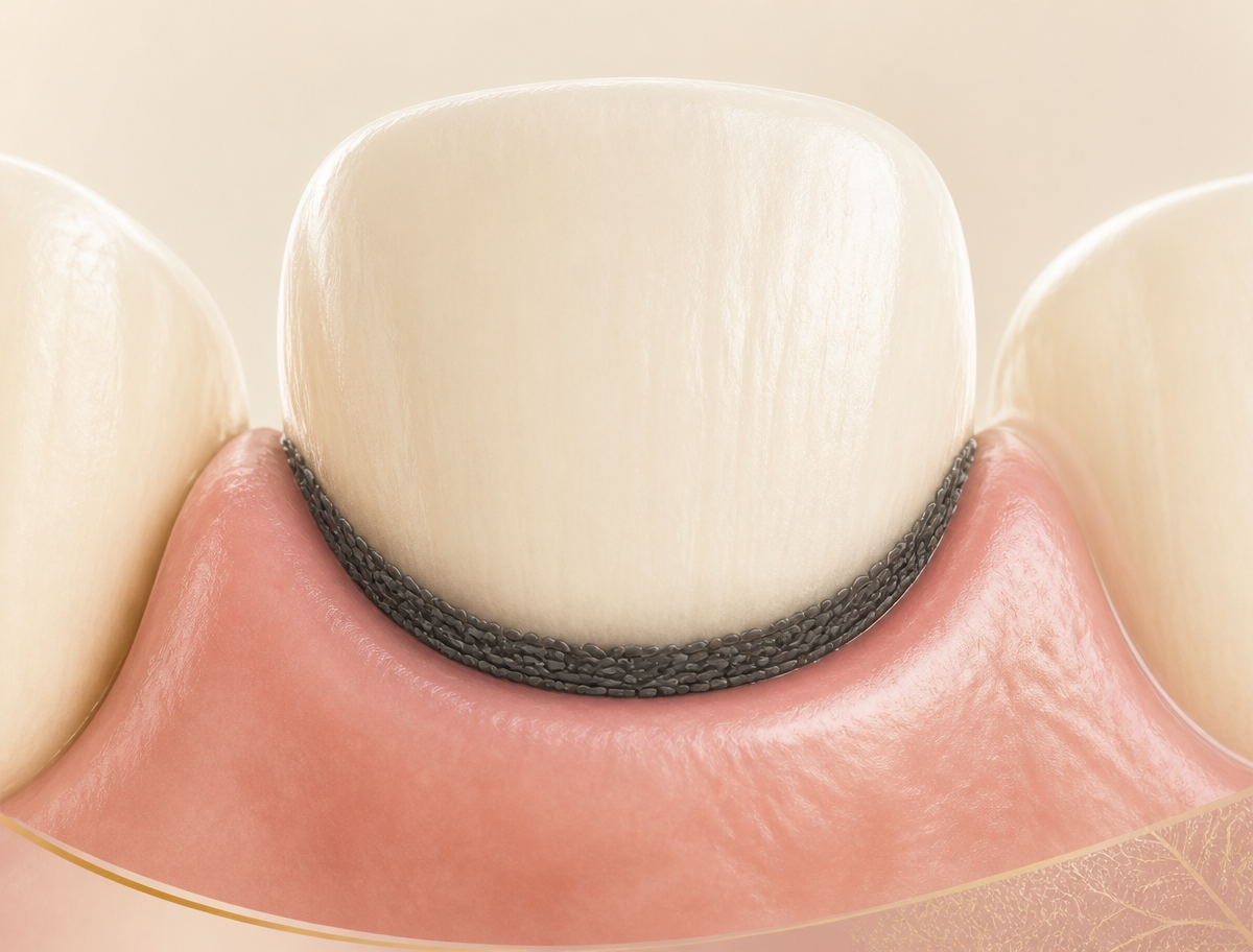
Not all extrinsic discoloration is deposited pigment. One distinct form — the dark line that follows the gum margin, often seen in children and in people with otherwise good hygiene — is bacterial in origin, produced by chromogenic organisms within the oral microbiome rather than by anything you ate or drank. It behaves differently from coffee staining in ways that matter: it returns after professional cleaning, it is associated with a distinguishable microbial profile, and the literature repeatedly reports it alongside a lower rather than higher caries rate — which is why a hygienist may tell you it is cosmetically annoying but not dangerous.
Peroxide chemistry does very little to it. Whitening gel oxidizes pigment molecules; it does not remove a bacterial deposit or change the community producing it. This kind of stain is removed mechanically and returns when the responsible organisms re-establish.
Whether the oral microbiome can be shifted to reduce this is an open research question. Probiotic approaches are being investigated and the early work is genuinely interesting, but the evidence base is small and should be read as preliminary, not settled.
Two layers, two different questions. The reason to understand both is that they answer different things. The oxidation chemistry on this page determines how much color you can recover. The film and the microbial community on your teeth determine how quickly that color comes back. Whitening well means working with both.
Ask ILUMI
Ask anything about whitening
Ask ILUMI is built on Dr. Steuer's clinical guidance and our complete reference FAQ. It knows the questions people ask most, from crowns and sensitivity to timing and aftercare, and answers in plain language, any hour of the day.
General information only — not dental advice. For decay, pain or extensive dental work, see your dentist.
What that looks like
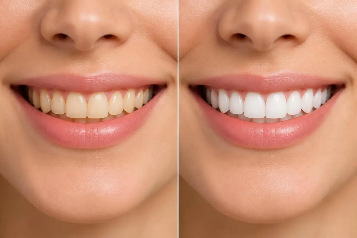
Shade change, in practice
Latest research
New from the ILUMI science desk
Whitening science
Why whitening makes teeth hurt — and what actually helps
Sensitivity is the main reason people abandon a whitening round. The mechanism is better understood than most product copy admits, and several of the standard fixes are weaker than they are sold as.
October 4, 2026 · 6 min read
Oral health
What actually stains your teeth — and one piece of advice that does not hold up
Tea outstains coffee. Tobacco's effect is measurable across a whole population. And the thing everyone tells you about brushing after acidic drinks has thinner evidence behind it than almost anyone admits.
October 4, 2026 · 6 min read
Whitening science
Why the light is two colors
Two wavelengths and a ten-minute session. The short session is doing more work than either light — here is what each part is actually for, including where the evidence does not support what this category usually claims.
August 30, 2026 · 7 min read
References
Where the mechanism comes from
These are the peer-reviewed and professional-body sources behind the mechanisms described on this page. They describe peroxide whitening and light activation generally; they are not studies of ILUMI products.
- [1] Allende JB, Campos-Bijit V, Morales-Gómez C, et al. Do laser and light activation improve in-office dental bleaching outcomes? An umbrella review of efficacy, tooth sensitivity, and biological safety. Journal of Esthetic and Restorative Dentistry. 2026;38(9):1665–1690.
Light activation did not consistently improve final whitening; photobiomodulation reduced post-bleaching sensitivity in placebo-controlled trials without impairing colour change. Effects and safety are parameter-dependent.
- [2] Maran BM, Burey A, de Paris Matos T, Loguercio AD, Reis A. In-office dental bleaching with light vs. without light: a systematic review and meta-analysis. Journal of Dentistry. 2018;70:1–13.
Light-activated in-office bleaching did not improve colour change compared with bleaching without light.
- [3] Carey CM. Tooth whitening: what we now know. Journal of Evidence-Based Dental Practice. 2014;14 Suppl:70–76.
Peroxide diffuses through enamel and dentin and oxidises pigmented organic molecules; the resulting smaller molecules reflect more light.
- [4] Kwon SR, Wertz PW. Review of the mechanism of tooth whitening. Journal of Esthetic and Restorative Dentistry. 2015;27(5):240–257.
Mechanism of hydrogen and carbamide peroxide bleaching, including the role of free-radical formation.
- [5] SoutoMaior JR, et al. Effectiveness of light sources on in-office dental bleaching: a systematic review and meta-analyses. Operative Dentistry. 2019;44(3):E105–E117.
Reviews the contribution of light activation to bleaching outcomes and to post-operative sensitivity.
- [6] Hanks CT, et al. / ADA Council on Scientific Affairs. Tooth whitening safety and efficacy statements.
American Dental Association overview of peroxide whitening safety, concentrations and sensitivity management.
- [7] Hamblin MR. Mechanisms and applications of the anti-inflammatory effects of photobiomodulation. AIMS Biophysics. 2017;4(3):337–361.
Red/near-infrared photobiomodulation and its documented anti-inflammatory and comfort effects in soft tissue.
- [8] Lendenmann U, Grogan J, Oppenheim FG. Saliva and dental pellicle — a review. Advances in Dental Research. 2000;14:22–28.
Formation and composition of the acquired enamel pellicle and its role as the interface between tooth surface and the oral environment.
- [9] Acquired salivary pellicle and oral diseases: a literature review. Journal of Taibah University Medical Sciences.
Protective functions of the salivary pellicle and its role in adsorption at the enamel surface.
- [10] Chromogenic bacterial staining of teeth: a scoping review. BMC Oral Health. 2025.
Scoping review of bacterially produced dental black stain, its microbial associations and its reported relationship to caries rate.
- [11] Al-Shareef et al. Current perspective on dental black stain of bacterial origin: a narrative review. European Journal of Oral Sciences. 2025.
Narrative review of the aetiology, recurrence and management of chromogenic black stain.
- [12] Probiotics to reduce microbiota-related dental stains: a potential approach.
Early-stage investigation of microbiome-directed approaches to chromogenic staining; preliminary evidence.
This page is educational and is not a substitute for individual dental advice. Consult your dentist before whitening if you have untreated decay, active gum disease, or extensive restorations on visible teeth.
Put the science to work
Dual-wavelength LED, a pH-balanced clinical gel, one 10-minute session a day.
Shop the system